What is our Five Component Oral Exam?
This is arguably the MOST important part of your horse’s routine dental care, as it allows us to systematically look at your horses head and mouth to evaluate functional efficiency and oral health to hopefully identify problems EARLY, so preventative measures can be taken.
Our five component oral check includes our external, oral soft tissue, occlusion, periodontal, and endodontic exams. This post will cover the external exam, with the other components being discussed in upcoming articles.
View all articles in our educational series.
The external exam
Lumps, and bumps, and swellings, oh my! We carefully look for any swellings or asymmetry to your horse’s face, as this can indicate an underlying problem.
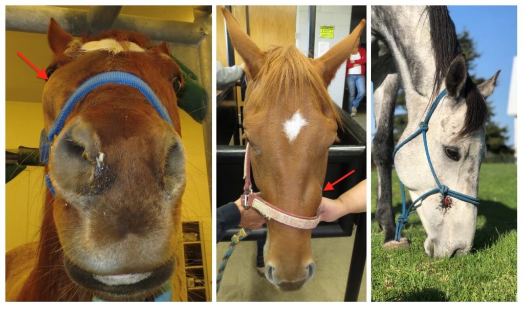
Not all of these are necessarily dental related. The horse on the left had a tumor in his sinus, creating a large facial swelling. The horse in the middle had an abscessed premolar that caused this large swelling. The horse on the right had a chronically abscessed tooth that caused a draining tract out the side of his face.
We also evaluate for any discharge from the eyes or nose. Again, these are not always related to a tooth problem, but sometimes can be!
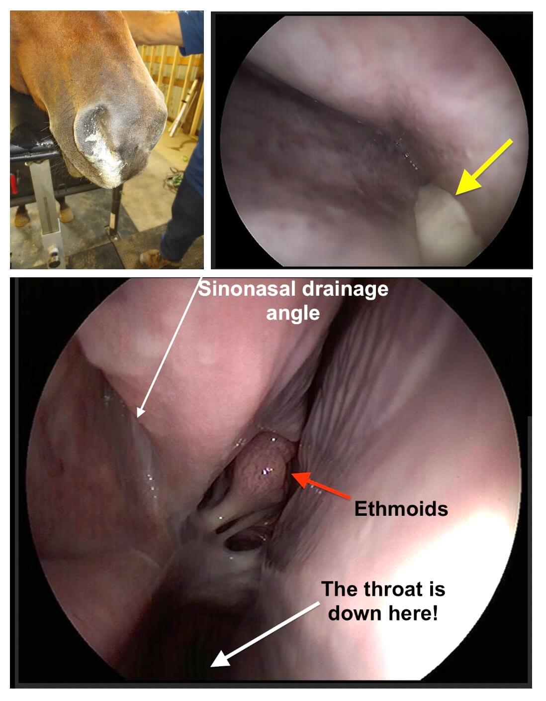
Here is an example of a horse with a sinus infection secondary to a tooth root abscess. The endoscopy picture shows drainage (yellow arrow) coming from the sino-nasal drainage angle.
It is also important to evaluate the muscles of mastication (chewing muscles!). Evidence of atrophy or asymmetry may indicate an underlying problem.
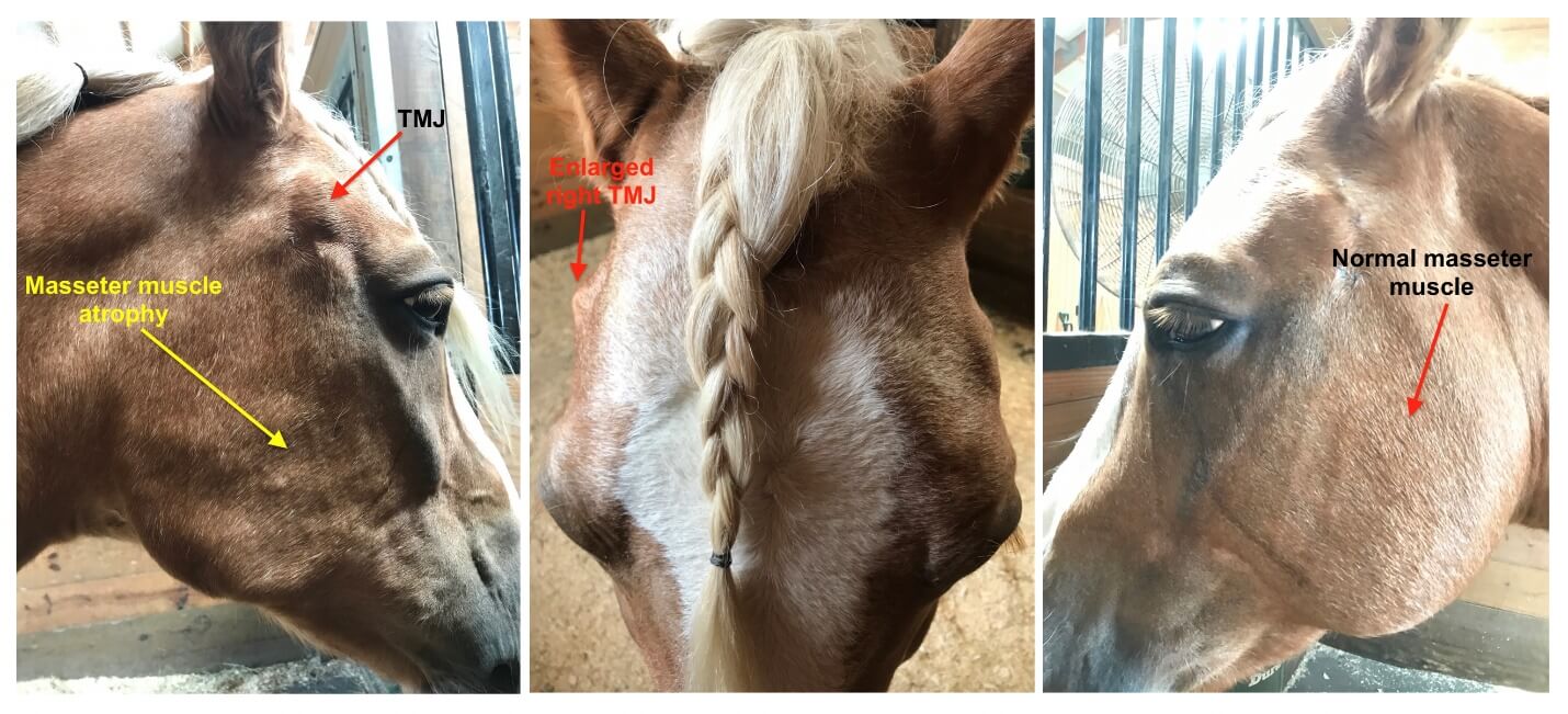
This horse developed atrophy of the masseter (cheek muscle) which is one of the primary muscles of chewing. In this horse’s case, the atrophy was only present on ONE side. You’ll notice that he had a slight enlargement of his right TMJ joint as well.
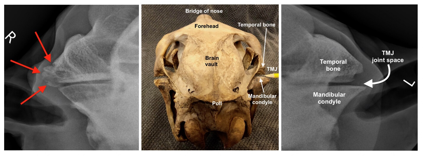
This horse required x-rays to further investigate the problem. You’ll see his x-rays below, with a skull specimen for reference. He has obvious arthritic changes present in his right temporomandibular joint (TMJ). This may have been from trauma at some point.
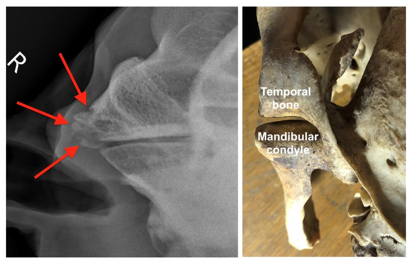
Here is another view of his affected TMJ next to a skull with a normal TMJ joint. You’ll notice the smooth margin on the normal joint, compared to the pitted and irregular surface of the bones on his x-ray. There are several treatment options available for horses with TMJ arthritis, including joint injections and surgical options to remove the mandibular condyle.
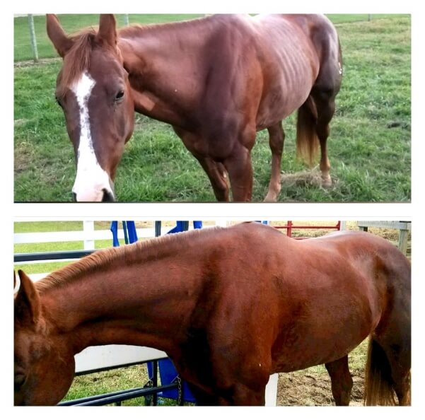
This picture shows a horse that had a septic TMJ that was resolved through surgical removal of his mandibular condyle. He was having significant difficulty chewing prior to surgery, as evidenced by his poor body condition. Six weeks after surgery, you will see he has bloomed back to a good body condition!