Our five component oral check includes our external, oral soft tissue, occlusion, periodontal, and endodontic exams. This post will cover endodontic status, with each of the other components being discussed in its own article.
View all articles in our educational series.
Endodontic status
The last component of the oral exam is the endodontic status. This comprises everything pertaining to the inside of the tooth — this includes the live part of the tooth and the tooth structure itself.
Endodontic disease can include anything from tooth cavities, tooth fracture, or tooth abscess. Read on to see examples of endodontic disease.
Warning: some pictures may have some blood.
Examples of endodontic disease
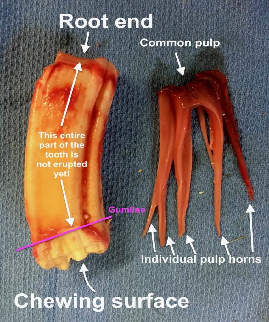
What is inside a tooth? The PULP! This is the “live” part of the tooth, and is made up of the blood supply, nervous tissue, and connective tissue.
In this picture, we have “plucked” the pulp out of this extracted tooth, which is from a young horse. When cheek teeth erupt into the mouth, they actually don’t have tooth roots yet.
You’ll notice on this tooth how much of the tooth actually sits below the gumline. The common pulp connects all of the finger-like pulp horns, which extend towards the tooth surface.
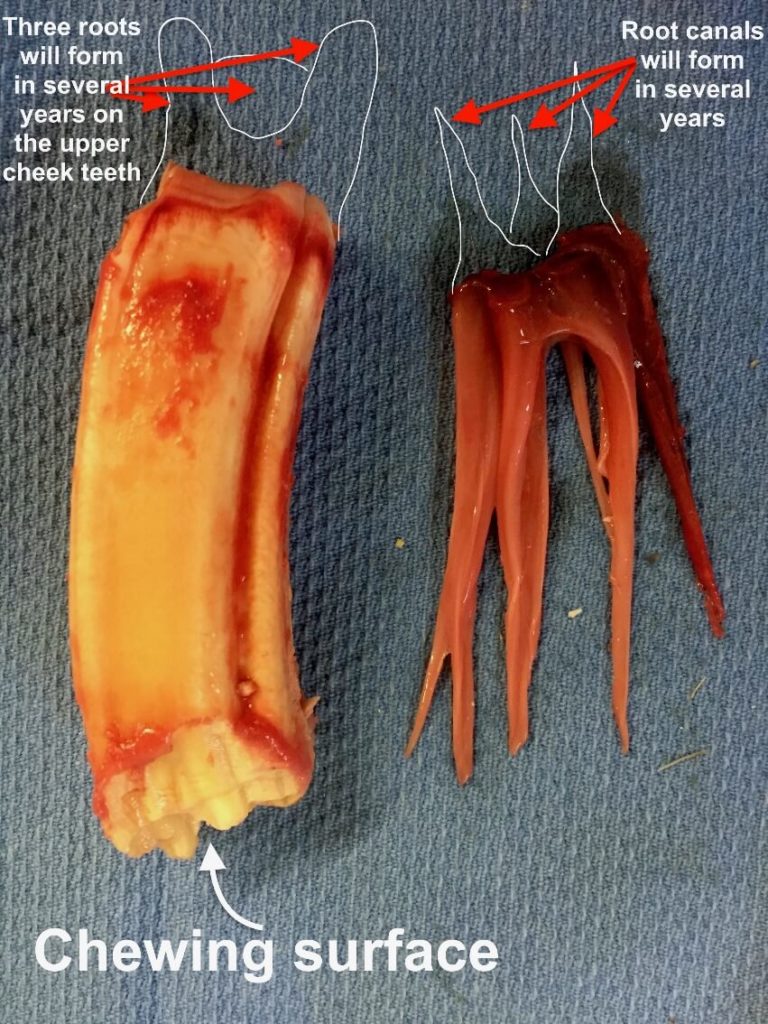
As the horse continues to age, eventually tooth root will form over the course of several years. You’ll see our representation of the tooth roots (three in this upper cheek tooth) as well as the corresponding root canals that form by branching off from the common pulp chamber.
As the horse continues to age, these pulp structures will get narrower and less robust, as they lay down secondary dentin to narrow down the pulp horns within the tooth.
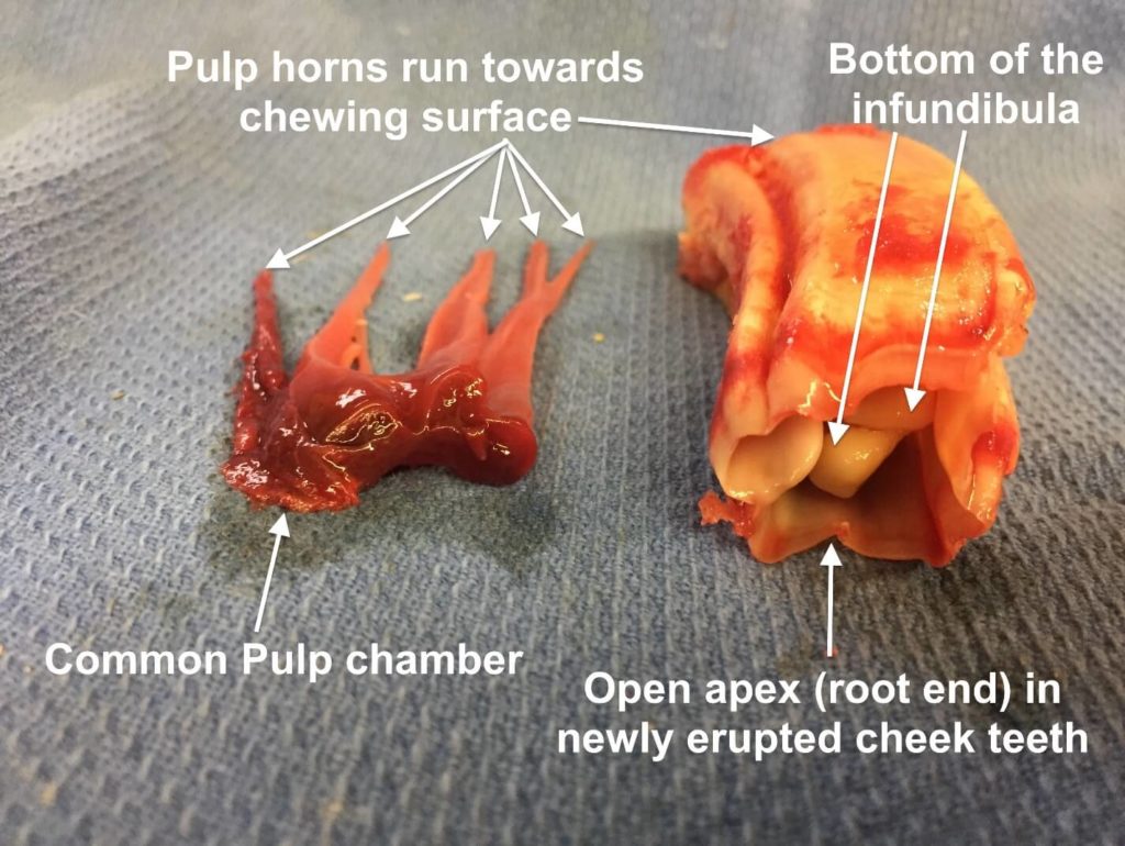
Here is another look at this tooth from a YOUNG horse, looking at the “root” end of the tooth. You’ll notice that it is completely wide open. This is why we were able to “pluck” the pulp out for demonstration purposes!
This also shows us the bottom of the “infundibula” which are enamel lined cups within the tooth. This information will make more sense when we start showing some of the endodontic problems that we may find on our oral exam.
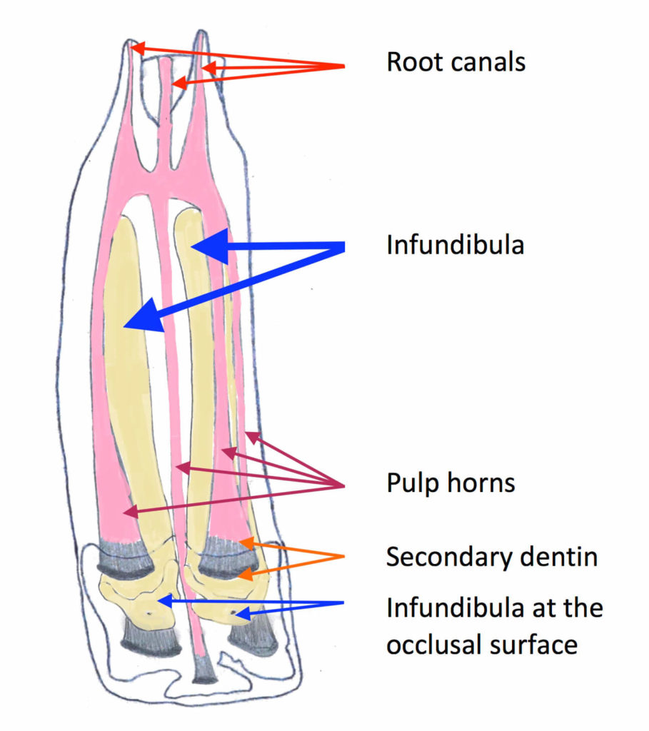
Here is one final depiction of what the inside of a tooth looks like, once the roots have formed.
The pink parts are the “pulp” which again, are the nerve and blood supply of the tooth. This live part is continuously depositing secondary dentin in the tips of the pulp horns so that the chewing surface of the tooth remains intact.
In the center of the tooth, you’ll notice the yellow INFUNDIBULA. These are enamel lined, inert structures within the center of the tooth. They are not connected to the nerve supply. They offer another ridge of enamel on the tooth surface for grinding food.
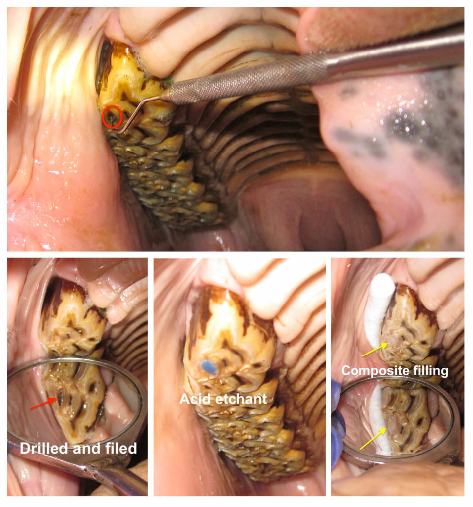
This is a horse that has pulp exposure evident on the oral exam. See where the probe is inserted into the tooth? (red circle) When we find this on our oral exam we always recommend taking an x-ray to find out if the tooth needs to be extracted or not.
In this case, we were able to offer a root canal type procedure, which you’ll see demonstrated in the bottom panel of pictures. We have followed this tooth for over 6 years, and it is still doing quite well!
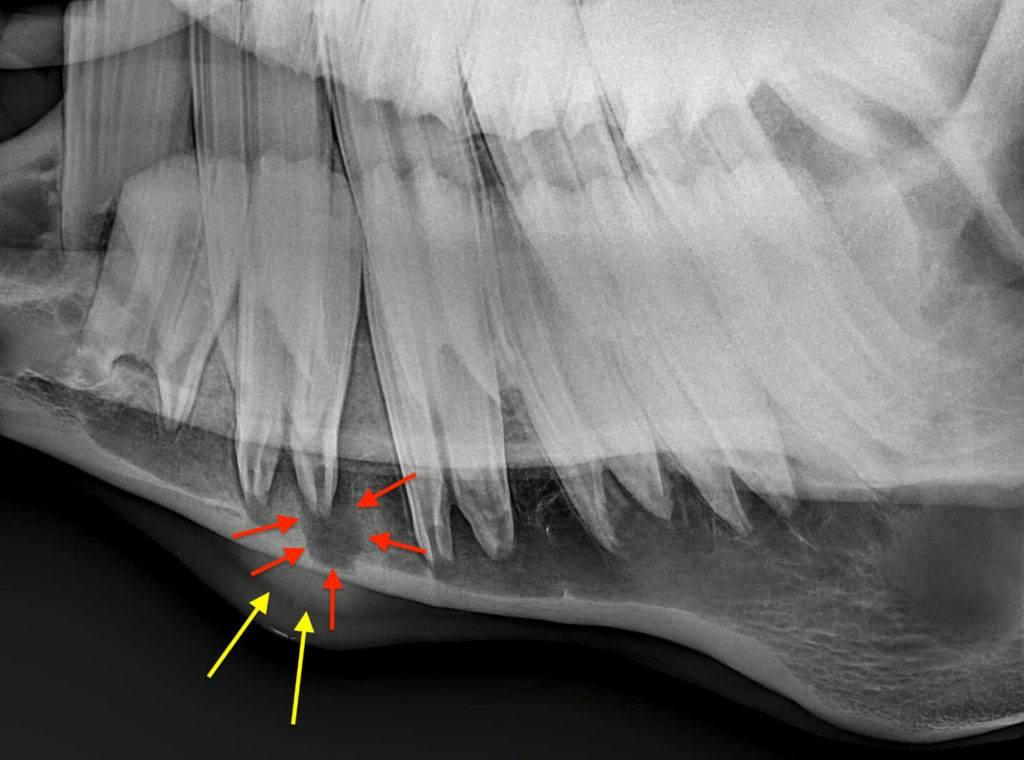
Here is an example of a horse with a tooth root abscess. You’ll notice the large halo around the root (red arrows).
In addition, this horse had a very large, firm swelling on its bottom jaw — notice the extra bone that has been laid down (yellow arrows) in response to this infection.
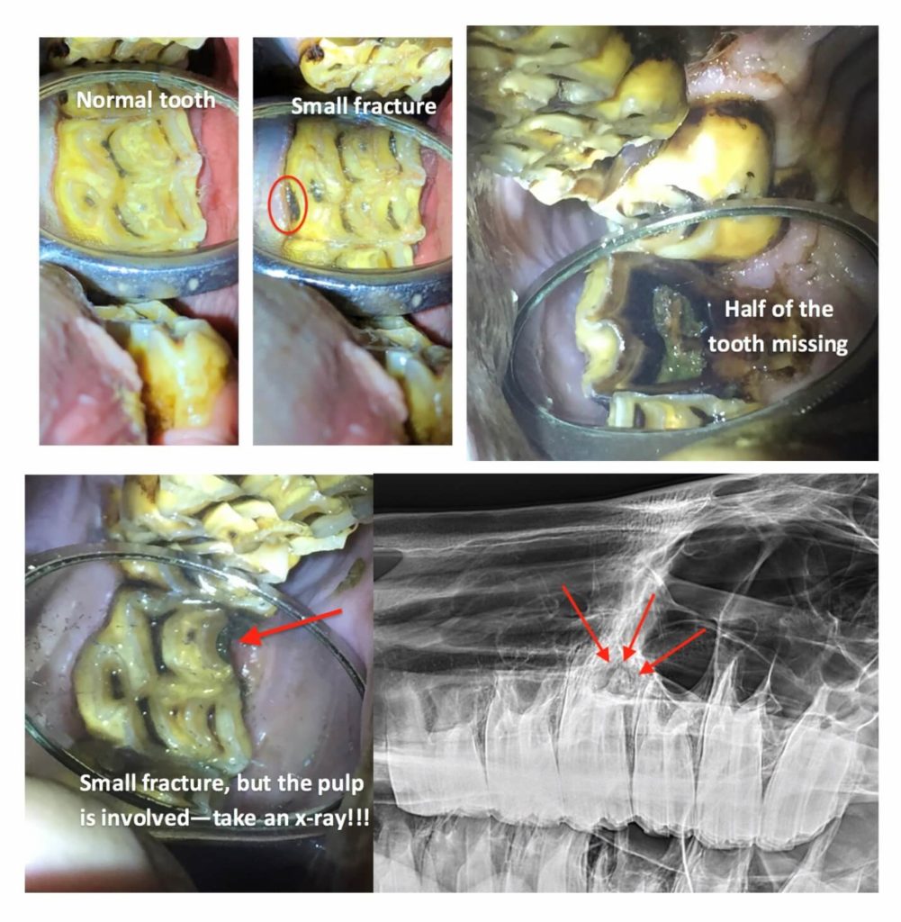
Not all fractures are created equally. The images in the top left show two relatively normal teeth.
The horse has fractured this tooth and half of the crown is missing. The horse in the bottom panes has a relatively small fracture evident on exam, however the pulp is involved in the fracture, so an x-ray must be taken to evaluate the tooth roots.
In this case, it was a good thing we did, because the root was abscessed (red arrows).
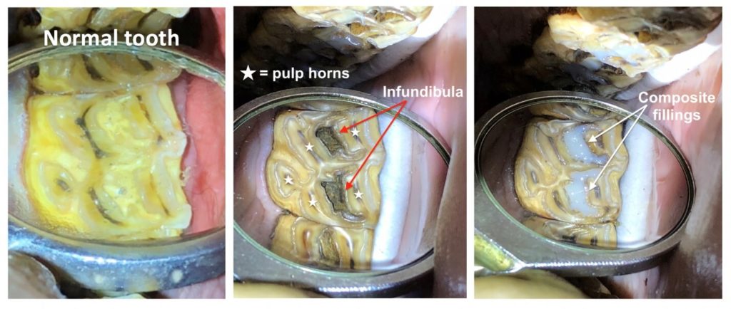
Some teeth get cavities, the most common location is in the infundibula. You’ll remember this is in the non-sensitive portion of the tooth. In some cases, depending on the age and level of decay present, we may restore these with fillings as we did in this horse.
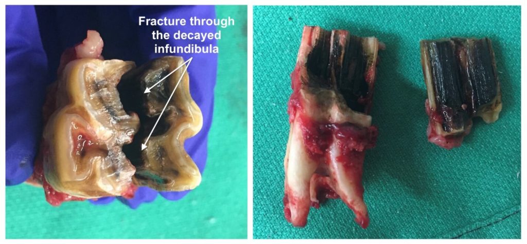
The reason why is important to monitor teeth with infundibular cavities (and sometimes restore them with fillings), is that in some cases, the tooth decay can result in tooth fracture.
This horse had a sinus infection related to this tooth fracture that resolved once the tooth was removed.